.webp&w=384&q=75)

Loading, please wait...
Loading, please wait...

Monitoring the health of a new organ is crucial for long-term success. Consequently, clinicians rely heavily on pediatric kidney transplant ultrasound to detect early complications. A recent longitudinal study from the Children's Medical Center (2018-2024) provides new insights into these imaging patterns. By analyzing over 1,000 scans from 164 patients, researchers identified which findings are transient and which signal potential graft failure.
The study revealed that nearly one-third of all postoperative ultrasounds appeared completely normal. However, abnormal findings were also frequent. Increased cortical echogenicity affected more than half of the participants. Moreover, approximately 30% of children developed perinephric fluid collections. Fortunately, these collections and abnormal resistive indices (RI) typically decreased as the graft matured. This trend suggests that many early irregularities are part of the normal healing process.
While some findings resolve, others require urgent attention. Specifically, the researchers found that hydronephrosis occurred in nearly 20% of the recipients. Unlike transient fluid collections, hydronephrosis significantly increased the risk of graft failure. Therefore, persistent dilation of the collecting system serves as a vital prognostic marker. Because of this, regular surveillance is necessary to ensure timely intervention for obstructive issues.
Furthermore, the study emphasizes the importance of longitudinal tracking rather than relying on a single scan. Since temporal trends show that most fluid collections dissipate, clinicians can often manage these conservatively. In contrast, any increase in resistive index or worsening hydronephrosis should prompt a more intensive investigation. Ultimately, these sonographic details help refine post-transplant care strategies in pediatric populations.
The most common findings include increased cortical echogenicity (52.4%) and perinephric fluid collections (29.9%). While many findings are transient, a normal scan occurs in about 32% of cases.
Yes, the study identified hydronephrosis as a significant risk factor for graft failure. It appeared in 19.5% of the patients and requires careful monitoring to protect the transplant.
Disclaimer: This content is for informational and educational purposes only and does not constitute medical advice or a professional relationship. Always seek the advice of a physician or other qualified health provider with any questions regarding a medical condition. Refer to the latest local and national guidelines for clinical practice.
References
Valizadeh P et al. Post-Transplant Ultrasound Findings in Pediatric Kidney Recipients: A Longitudinal Experience From the Children's Medical Center (2018-2024). J Clin Ultrasound. 2026 Mar 17. doi: 10.1002/jcu.70229. PMID: 41844519.
Franke D. The diagnostic value of Doppler ultrasonography after pediatric kidney transplantation. Pediatr Nephrol. 2022;37(6):1229-1240. doi:10.1007/s00467-021-05253-y.
Nixon GW et al. Imaging of Pediatric Renal Transplants and Their Complications: A Pictorial Review. RadioGraphics. 2013;33(5):1227-1251. doi:10.1148/rg.335125150.

Read summarized clinical updates, watch expert medical content, and earn CME certifications right from your smartphone.
Open your smartphone camera to scan and install.





Longitudinal study analyzing ultrasound findings in 164 pediatric kidney recipients, highlighting hydronephrosis as a key predictor of graft failure....
7 months ago

A retrospective cohort study reveals that frailty quadruples the odds of pressure injuries in hospitalized older adults. Integrating the Clinical Frailty Scale with comprehensive geriatric care is crucial for identifying vulnerability early and implementing targeted multidisciplinary prevention.
Today

Occult maternal malignancy accounts for 3.82% of persistent unreportable NIPT results with genomic noise. Multiple chromosomal aberrations, notably on chromosome 18, warrant thorough oncological evaluation and whole-body MRI to ensure timely maternal diagnosis while preserving fetal care.
Today

This article reviews the forensic and clinical complexities of acute myocarditis arising on left ventricular hypertrophy in children, highlighting early diagnostic red flags and multidisciplinary postmortem autopsy protocols.
Today
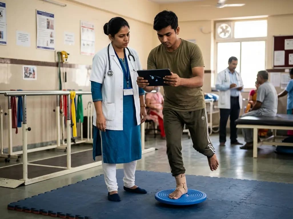
A randomized controlled trial demonstrates that an eight-week dual-task training intervention significantly improves static and dynamic postural control over standard balance exercises in individuals with chronic ankle instability, addressing critical neurocognitive deficits during athletic and daily activities.
Today

A retrospective cohort study of 7,716 ICU patients from the MIMIC-IV database demonstrates that Gradient Boosting Decision Trees significantly outperform traditional SOFA scores in predicting in-hospital mortality among patients with sepsis and diabetes, with SHAP analysis providing clinical interpretability.
Today
