
Loading, please wait...
Loading, please wait...

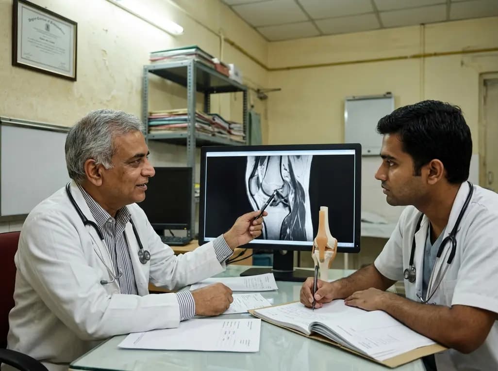
Magnetic resonance imaging (MRI) has revolutionized diagnostic medicine by providing detailed anatomical images. However, a new frontier is emerging through MRI conductivity imaging. This noninvasive technique allows clinicians to probe the electrical properties of biological tissues, specifically electrical conductivity and relative permittivity. By analyzing these parameters, doctors can gain deeper insights into tissue microstructure and ionic composition that standard imaging often misses.
The clinical utility of this technology depends heavily on the frequency regime used during scanning. At low frequencies, typically below 1 MHz, conductivity mapping primarily reflects the underlying tissue microstructure. Conversely, at high frequencies around 100 MHz, the maps primarily highlight changes in ionic composition. Consequently, these variations provide unique biomarkers for different pathological states, ranging from inflammation to malignancy.
Research highlights significant potential for MRI conductivity imaging in oncology and neurology. In cancer care, tumor tissues often exhibit higher conductivity due to increased water content and shifted ion concentrations. This allows radiologists to distinguish between benign and malignant lesions more accurately. Furthermore, clinicians can use these maps to monitor how tumors respond to chemotherapy or radiation in real-time. This capability improves therapeutic monitoring and helps in adjusting treatment plans early.
In neurology, conductivity imaging shows promise in identifying subtle changes in brain tissue. Specifically, it helps in detecting early signs of neurodegeneration or ischemia. Because the technique uses standard MRI hardware, integrating it into routine clinical protocols is increasingly feasible. Moreover, technical developments in reconstruction algorithms like electrical properties tomography (EPT) continue to enhance image quality and diagnostic reliability.
While challenges such as signal-to-noise ratios and reconstruction artifacts remain, the field is advancing rapidly. Integrating these electrical property maps into routine practice could significantly improve diagnostic precision. Ultimately, this technology offers a multi-parametric approach to patient care, combining traditional anatomy with functional electrical data.
Standard MRI focuses on proton density and relaxation times to create anatomical images. In contrast, conductivity imaging measures how tissues conduct electricity, providing information about ion concentration and cellular structure that traditional scans cannot see.
Yes, most methods like Electrical Properties Tomography (EPT) are entirely noninvasive and do not require external current injection. They utilize the radiofrequency fields already present during a standard MRI session, making them safe for clinical use.
Disclaimer: This content is for informational and educational purposes only. It does not constitute medical advice, diagnosis, or treatment. Always seek the advice of a qualified healthcare provider with any questions regarding a medical condition. Refer to the latest local and national guidelines for clinical practice.
References
Mandija S et al. Clinical Applications of Electrical Conductivity Imaging Using MRI. J Magn Reson Imaging. 2026 Mar 07. doi: 10.1002/jmri.70279. PMID: 41795132.
Jahng GH, et al. Principle, Development, and Application of Electrical Conductivity Mapping Using Magnetic Resonance Imaging. Progress in Medical Physics. 2024;35(4):73-85. doi: 10.14316/pmp.2024.35.4.73.
Zhang X, et al. Magnetic-resonance-based electrical properties tomography: a review. IEEE Reviews in Biomedical Engineering. 2014;7:87-96. doi: 10.1109/RBME.2013.2297206.

Read summarized clinical updates, watch expert medical content, and earn CME certifications right from your smartphone.
Open your smartphone camera to scan and install.


Explore the emerging role of electrical conductivity imaging in MRI for better tissue characterization and diagnostic precision in clinical settings....
4 months ago

Andhra Pradesh reported 10 new Covid-19 cases, taking the state tally to 49 while deaths remain at four. With 24 patients hospitalized and 16 under home isolation, the Health Department has intensified monitoring. Medical professionals should review regional distribution, diagnostic protocols, and management plans.
Today

An 11-year Swedish registry study of 618 uterine sarcoma patients found that minimally invasive surgery yielded survival comparable to open surgery in early stages. However, adjuvant chemotherapy conferred no survival benefit in localized or advanced disease, highlighting stage and histology as key outcomes.
3 days back

A cross-sectional study evaluates post-intensive care syndrome in cardiac patients 2-4 weeks post-ICU discharge, highlighting cognitive, psychological, and functional impairments and the need for structured multidisciplinary rehabilitation.
3 days back
Anterior cruciate ligament reconstruction failure lacks uniform definition. A narrative review proposes an integrative framework incorporating objective and subjective instability, persistent pain, restricted motion, graft rupture, and secondary meniscal injury to standardize clinical reporting.
3 days back

With World Obesity Atlas data warning that over 41 million Indian children are overweight or obese, ICMR and NIN have unveiled a 10-point policy roadmap. The initiative calls for mandatory front-of-pack labeling, HFSS taxes, strict marketing bans, and healthier school environments to curb non-communicable diseases.
Today
