
Loading, please wait...
Loading, please wait...

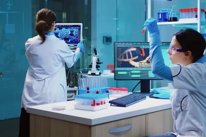
Prenatal diagnosis of rare vascular conditions such as fetal dural sinus malformations (DSM) remains a significant challenge for obstetricians and radiologists. These anomalies involve the abnormal dilation of the dural sinuses, often affecting the torcular Herophili. While traditional 2D ultrasound provides a baseline for detection, its ability to delineate complex venous anatomy is sometimes limited. Therefore, incorporating three-dimensional (3D) imaging techniques has become essential for comprehensive fetal assessment and parent counseling.
Recent clinical evidence suggests that 3D ultrasound significantly improves the characterization of fetal dural sinus malformations. Unlike 2D imaging, 3D technology allows clinicians to visualize venous continuity and the precise morphology of intraluminal thrombi. Furthermore, 3D ultrasound helps confirm the absence of arterialization, which is a critical step in differentiating DSM from other arteriovenous shunts. Consequently, the integration of these advanced imaging modalities into routine protocols offers a more detailed anatomical map of the fetal brain.
In a retrospective study of eight fetuses, researchers identified various sonographic markers that correlate with postnatal health. Lesions often involved the torcular Herophili and superior sagittal sinus, sometimes reaching sizes up to 90 × 110 mm. Moreover, the presence of thrombosis, found in over 60% of cases, served as a key indicator of adverse outcomes. Despite the severity of some cases, nearly 50% of the liveborn infants achieved favorable neurodevelopmental milestones. These findings highlight that while the diagnosis is serious, meticulous prenatal monitoring can lead to successful management in many instances.
Clinicians should prioritize combined 2D and 3D ultrasound whenever they suspect a midline cranial anomaly. Because 3D imaging offers superior lesion delineation, it enables more accurate risk stratification. Additionally, genetic testing remains a vital part of the workup, although many cases of DSM show normal chromosomal results. By using a multi-modal approach, medical teams can better predict potential complications like hydrocephalus or fetal growth restriction, ultimately improving the quality of perinatal care.
Diagnosis typically relies on identifying cystic or anechoic lesions near the dural sinuses. Specifically, 2D ultrasound often detects these masses in the posterior fossa, while 3D ultrasound clarifies the presence of thrombosis and venous connections.
No, the prognosis varies. Although some cases involve fetal demise or the need for pregnancy termination, many children show favorable neurodevelopmental outcomes. The presence of thrombosis and associated anomalies like hydrocephalus are the most critical prognostic indicators.
3D ultrasound provides better spatial resolution and the ability to view the lesion in multiple planes simultaneously. Consequently, it allows for a more thorough evaluation of the thrombus morphology and the surrounding vascular architecture compared to 2D alone.
Disclaimer: This content is for informational and educational purposes only. It does not constitute professional medical advice, diagnosis, or treatment. Always seek the advice of your physician or other qualified health provider with any questions you may have regarding a medical condition. Refer to the latest local and national guidelines for clinical practice.
References

Read summarized clinical updates, watch expert medical content, and earn CME certifications right from your smartphone.
Open your smartphone camera to scan and install.


A new case series highlights how combining 2D and 3D ultrasound improves the detection and prognostic assessment of fetal dural sinus malformations....
4 months ago

A recent cross-sectional study of 15.2 million adults reveals a goiter prevalence of 1.3-2.5%. This detailed analysis explores why women and older adults are at higher risk, the dominance of non-toxic multinodular subtypes, and the clinical implications for primary care and endocrinology.
Today
New research proposes a three-tier biomechanical framework for managing cesarean-related scars. While superficial treatments like silicone and taping are well-supported, direct mechanotherapy for uterine niches remains a hypothesis-generating area requiring further clinical validation.
Today
This case study examines a 56-year-old man who experienced sequential bilateral common carotid artery occlusion. Findings revealed protein C deficiency and a patent foramen ovale (PFO), suggesting paradoxical embolism as the primary cause. This highlights the need for thorough thrombophilia screening.
Today
This case study examines the first MRI-documented instance of indirect decompression following a single-position robot-assisted lateral lumbar corpectomy for a traumatic L4 burst fracture, demonstrating significant canal restoration and reduced surgical morbidity.
Today
An international panel of experts has officially recognized fatty pancreas disorder as a distinct disease in the Melbourne Consensus. Learn about its diagnostic criteria using MRI, its pathophysiological association with type 2 diabetes and cancer, and evidence-based lifestyle and pharmacological interventions.
Today
