
Loading, please wait...
Loading, please wait...

Thyroid carcinoma incidence continues to rise globally, making precise preoperative assessment a critical clinical priority. Specifically, identifying capsular invasion and neural invasion (NI) before surgery is essential because these factors determine patient recurrence and survival rates. Traditionally, radiologists have found it difficult to detect these subtle invasive features using conventional imaging alone. However, recent research highlights how CT radiomics thyroid carcinoma machine learning models provide a robust, noninvasive solution for preoperative risk stratification.
Radiomics transforms medical images into high-dimensional, minable data. Consequently, clinicians can extract quantitative features that the human eye might miss. In a retrospective cohort of 111 patients, researchers extracted 111 gray-level co-occurrence matrix features from arterial and venous phase CT scans. Notably, the team selected nine key radiomic features using least absolute shrinkage and selection operator regression. This process ensures that the physical meaning of texture features, such as tumor microstructural heterogeneity, remains preserved.
The study evaluated several diagnostic approaches, including clinical nomograms, random forest (RF) models, and neural networks (NN). Furthermore, the clinical indicator-based nomogram achieved an impressive Area Under the Curve (AUC) of 0.9418 for predicting capsular invasion. Meanwhile, the radiomic-based nomogram also performed strongly, showing an AUC of 0.9334. These results suggest that integrating digital biomarkers with clinical data significantly improves diagnostic accuracy. Therefore, surgeons can use these tools to tailor their approach, potentially reducing the need for aggressive re-operations.
Accurate prediction of neural invasion is vital for maintaining a patient's quality of life after surgery. The multimodal neural network model showed promising stability in identifying NI risk. Consequently, this technology allows for a more personalized treatment plan for patients in India and worldwide. Moreover, the stability of these models was verified using 5-fold cross-validation and bootstrap resampling. By identifying high-risk patients preoperatively, oncologists can ensure more aggressive monitoring or targeted therapies from the outset.
Radiomics uses advanced algorithms to analyze pixel-level data, revealing tumor patterns like heterogeneity that are invisible to the human eye. This provides a more objective and quantitative assessment of tumor aggressiveness than visual inspection alone.
Neural invasion is a pivotal prognostic factor that correlates with higher recurrence rates. Preoperative detection helps surgeons plan nerve-sparing techniques or decide the extent of resection needed to achieve clear margins.
While these models show high diagnostic accuracy, they currently serve as supportive tools. Clinicians should integrate radiomic data with clinical indicators, such as galectin-3 levels, to make the most informed treatment decisions.
Disclaimer: This content is for informational and educational purposes only. It does not constitute medical advice and should not replace professional consultation. Refer to the latest local and national guidelines for clinical practice.
References
1. Cong FF et al. CT Radiomics-Based Machine Learning Model for Predicting Capsular and Neural Invasion in Thyroid Carcinoma: Diagnostic Accuracy Study. JMIR Med Inform. 2026 Mar 12. doi: 10.2196/77349. PMID: 41818775.
2. Yu P et al. Radiomics Analysis of Computed Tomography for Prediction of Thyroid Capsule Invasion in Papillary Thyroid Carcinoma: A Multi-Classifier and Two-Center Study. Front Oncol. 2023;13:1134069.
3. Bhat S et al. Predictive Modelling Using Thyroid Cartilage Segmentation and Radiomic Features: A Feasibility Study. Int J Otolaryngol Head Neck Surg. 2025; DOI: 10.1007/s12070-025-05609-y.
"
Read summarized clinical updates, watch expert medical content, and earn CME certifications right from your smartphone.
Open your smartphone camera to scan and install.


A new study demonstrates that machine learning models using CT radiomics can accurately predict capsular and neural invasion in thyroid carcinoma patients....
4 months ago
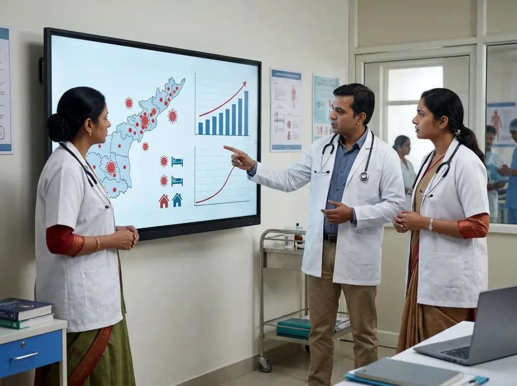
Andhra Pradesh reported 10 new Covid-19 cases, taking the state tally to 49 while deaths remain at four. With 24 patients hospitalized and 16 under home isolation, the Health Department has intensified monitoring. Medical professionals should review regional distribution, diagnostic protocols, and management plans.
Today

An 11-year Swedish registry study of 618 uterine sarcoma patients found that minimally invasive surgery yielded survival comparable to open surgery in early stages. However, adjuvant chemotherapy conferred no survival benefit in localized or advanced disease, highlighting stage and histology as key outcomes.
3 days back
A cross-sectional study evaluates post-intensive care syndrome in cardiac patients 2-4 weeks post-ICU discharge, highlighting cognitive, psychological, and functional impairments and the need for structured multidisciplinary rehabilitation.
3 days back
Anterior cruciate ligament reconstruction failure lacks uniform definition. A narrative review proposes an integrative framework incorporating objective and subjective instability, persistent pain, restricted motion, graft rupture, and secondary meniscal injury to standardize clinical reporting.
3 days back

With World Obesity Atlas data warning that over 41 million Indian children are overweight or obese, ICMR and NIN have unveiled a 10-point policy roadmap. The initiative calls for mandatory front-of-pack labeling, HFSS taxes, strict marketing bans, and healthier school environments to curb non-communicable diseases.
Today
