.webp&w=384&q=75)

Loading, please wait...
Loading, please wait...

Colorectal liver metastases (CRLM) present a significant hurdle in clinical practice because outcomes after surgery vary extensively. Currently, surgeons rely on clinical risk scores to plan treatment, but these markers often fail to capture the full biological complexity of the tumor. Consequently, researchers are turning to a CT radiomics signature to provide more precise, non-invasive prognostic information.
A recent study published in Physics in Medicine & Biology utilized preoperative CT scans from 197 patients to develop a predictive model. The team extracted 851 radiomics features and 256 deep features from convolutional neural networks. By applying advanced statistical methods, they identified an eight-feature signature that generates a specific risk score, known as the Rad score.
The CT radiomics signature demonstrated impressive accuracy in identifying patients at high risk of poor survival. In the training cohort, the Rad score emerged as a powerful independent predictor of overall survival. Specifically, the high-risk group showed a hazard ratio of 2.671 compared to the low-risk group. Moreover, the model maintained its predictive power across various timeframes, achieving Area Under the Curve (AUC) values of 0.781 at one year and 0.712 at five years.
Furthermore, this imaging biomarker significantly outperformed traditional clinical variables. While standard clinical risk factors provide a baseline, integrating the radiomics-based Rad score allows for more granular stratification. This breakthrough suggests that oncologists can better identify which patients may require more aggressive adjuvant therapies or closer post-operative monitoring.
Additionally, the study highlights the potential of combining deep learning with traditional radiomics. By processing both human-engineered features and deep neural network insights, the model captures tumor heterogeneity that is invisible to the naked eye. This transition toward digital pathology and imaging ensures that surgical planning becomes increasingly personalized.
A CT radiomics signature is a combination of quantitative data points extracted from medical images. These features describe tumor texture, shape, and intensity, providing a digital fingerprint of the cancer's biology.
It helps clinicians by providing a non-invasive way to predict how long a patient might survive after surgery. This allows for better risk stratification, helping doctors decide which patients are most likely to benefit from resection.
Rather than replacing them, this signature complements existing scores. It provides a higher level of accuracy when combined with clinical data, leading to more informed decision-making in oncology.
Disclaimer: This content is for informational and educational purposes only. It does not constitute medical advice or a professional relationship. Always consult a qualified healthcare provider for diagnosis and treatment. Refer to the latest local and national guidelines for clinical practice.
References
Kha QH et al. A CT radiomics signature enables risk stratification and survival prediction in colorectal liver metastases. Phys Med Biol. 2026 Mar 02. doi: 10.1088/1361-6560/ae4c14. PMID: 41771176.
Wang A et al. Exploring tumor heterogeneity in colorectal liver metastases by imaging: Unsupervised machine learning of preoperative CT radiomics features for prognostic stratification. Eur J Radiol. 2024 Apr;173:111459. doi: 10.1016/j.ejrad.2024.111459.
Deng Y et al. Predicting one-year post-surgical recurrence in colorectal liver metastasis using CT radiomics and machine learning. PLOS ONE. 2025 Aug 29;20(8):e0329481. doi: 10.1371/journal.pone.0329481.
"
Read summarized clinical updates, watch expert medical content, and earn CME certifications right from your smartphone.
Open your smartphone camera to scan and install.





Researchers have developed a CT radiomics signature that provides non-invasive risk stratification and survival prediction for colorectal liver metastases....
7 months ago
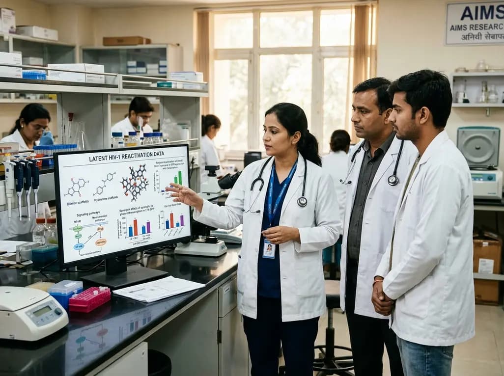
Novel diimide and hydrazone scaffolds, particularly compound #C4, synergistically reactivate latent HIV-1 when combined with JQ1 or PKC activators. Demonstrating donor-matched proviral transcription ex vivo and non-canonical signaling, these findings advance shock and kill strategies toward reservoir eradication.
Today

A cross-sectional study reveals that maternal RSV vaccination coverage reached only 43.7% in Argentina, with maternal vaccine knowledge being the sole independent predictor of uptake. Clinicians play a crucial role in eliminating informational and structural barriers to optimize infant health.
Today

Review how tailored exercise programs benefit frail older patients with heart failure. Explore pathophysiological mechanisms, individualized training prescriptions, and strategies to overcome adherence barriers in clinical practice.
Yesterday

This comparative study evaluates posterior labral tears with and without glenohumeral instability, highlighting distinct demographic patterns, MRI underestimation in stable shoulders, and successful arthroscopic repair outcomes.
Yesterday

A clinical study demonstrates that early intervention with tocilizumab, either as monotherapy or bridged with corticosteroids, achieves significant proptosis reduction and resolves active inflammation in treatment-naive thyroid eye disease.
Yesterday
