.webp&w=384&q=75)

Loading, please wait...
Loading, please wait...
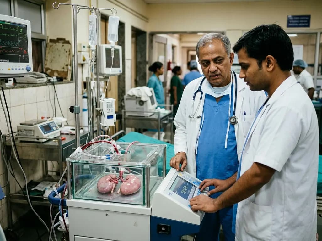
Expanding the deceased donor pool remains a critical goal in modern solid organ transplantation. Donation after circulatory death (DCD) provides valuable donor organs, but these grafts endure prolonged agonal and warm ischemic phases. Consequently, ischemia-reperfusion injury presents a formidable barrier to post-transplant graft survival. To overcome these logistical and biological challenges, clinicians are investigating en bloc kidney HOPE as a pragmatic rescue technique. This preservation approach delivers continuous oxygenation under cold conditions, stabilizing compromised renal parenchyma before implantation.
Transplant surgeons frequently encounter severe physiological compromise when retrieving kidneys from circulatory death donors. Because donor warm ischemia begins during the agonal phase prior to cardiac arrest, renal parenchymal cells rapidly lose intracellular adenosine triphosphate. Consequently, cellular ion pumps fail, leading to intracellular sodium accumulation, mitochondrial swelling, and endothelial damage. When teams preserve these fragile organs using traditional static cold storage, hypothermia alone fails to halt progressive metabolic deterioration. Furthermore, sudden rewarming and reperfusion in the recipient trigger reactive oxygen species production, complement activation, and severe microvascular spasm.
To interrupt this pathological cascade, clinicians increasingly implement hypothermic oxygenated machine perfusion. Supplying oxygen at low temperatures sustains minimal mitochondrial respiration without generating harmful free radicals. Therefore, mitochondria recharge their energetic potential and replenish adenine nucleotide pools before graft rewarming. In high-risk scenarios, en bloc kidney HOPE provides continuous active metabolic support to both kidneys concurrently. This method fundamentally shifts organ preservation from passive hypothermic storage to active metabolic resuscitation. As a result, transplant recipients experience significantly lower rates of delayed graft function.
Standard renal machine perfusion typically requires individual vascular cannulation of each renal artery. However, this conventional approach poses significant logistical challenges when anatomical anomalies or extensive vascular plaques complicate back-table preparation. Furthermore, repeated manipulation of delicate renal arteries increases the risk of intimal dissection, vascular spasm, and mechanical thrombosis. To circumvent these hazards, surgeons can retrieve both kidneys en bloc with an intact segment of the donor abdominal aorta and inferior vena cava.
During back-table preparation, the surgical team ligates lumbar branches and oversews the proximal aortic stump. Subsequently, clinicians cannulate the distal aortic conduit, creating a secure, high-volume inflow tract. This arrangement delivers perfusate evenly to both renal vascular beds under uniform hydrostatic pressure. In addition, the inferior vena cava acts as an uninhibited outflow drainage channel, preventing venous congestion and parenchymal oedema. Therefore, en bloc aortic perfusion eliminates delicate microvascular cannulation while preserving vascular integrity. This streamlined approach saves critical minutes during organ retrieval and establishes an exceptionally stable interface between the organ bloc and the perfusion circuit.
Specialized single-organ perfusion machines and their corresponding disposables are not always immediately available across every transplant centre. When equipment shortages threaten organ viability, transplant teams can utilize flexible multiorgan preservation platforms such as the VitaSmart system. Surgeons can configure these multiorgan consoles to operate safely under renal-specific parameters. Specifically, the machine maintains low hydrostatic pressures between twenty and thirty millimetres of mercury, which protects delicate glomerular capillaries from shear stress.
Simultaneously, the closed circuit circulates a cold, oxygen-saturated preservation solution across the renal vascular network. Ultrasonic flow sensors and inline pressure transducers deliver continuous, real-time hemodynamic feedback to the clinical team. Consequently, surgeons can observe changes in flow dynamics and calculate vascular resistance indexes over time. An unexpected rise in resistance warns clinicians of potential microvascular thrombosis or vasospasm, whereas a progressive decline indicates stable microcirculatory filling. Moreover, access ports permit timely pharmacological supplementation, including vasodilators or cytoprotective agents. Thus, repurposing multiorgan consoles provides an adaptable, cost-effective solution without compromising perfusion quality.
A profound advantage of hypothermic oxygenated perfusion lies in its capacity for dynamic, objective graft evaluation. Traditional static cold storage leaves clinicians blind to true parenchymal viability until surgical revascularization occurs in the recipient operating room. In contrast, continuous machine perfusion acts as a biological stress test, revealing critical data regarding microvascular resistance and metabolic competence. During prolonged perfusion, surgeons monitor vascular flow trends; stable flow at low pressure reliably correlates with intact capillary beds.
Furthermore, serial perfusate sampling allows laboratory quantification of ischemic injury biomarkers. Clinicians routinely evaluate flavin mononucleotide release from mitochondrial complex I, which strongly predicts post-transplant mitochondrial dysfunction and graft failure. In addition, teams measure lactate dehydrogenase, lactate accumulation, and perfusate acid-base status. When these metabolic indicators remain within acceptable thresholds over extended perfusion intervals, transplant surgeons can confidently accept borderline organs that they might otherwise discard. Therefore, real-time biomarker profiling transforms organ evaluation from subjective anatomical judgment into an objective assessment of tissue vitality, protecting recipients from primary non-function.
Organ transplantation frequently demands extraordinary logistical coordination across distant medical centres. Recipient preparation, crossmatch verification, and surgical theatre scheduling frequently induce unavoidable delays that extend cold ischemia time. In deceased circulatory donation, every additional hour of static cold storage exacerbates subsequent ischemia-reperfusion injury. However, recent feasibility data demonstrate that kidneys can safely undergo eighteen hours of continuous hypothermic oxygenated perfusion without structural degradation or functional decline.
Because active oxygenation continuously recharges mitochondrial reserves, extended machine perfusion effectively decouples organ retrieval time from recipient surgery timing. Consequently, surgical teams can convert stressful emergency night-time procedures into controlled daytime operations without jeopardizing graft survival. Furthermore, prolonged preservation windows allow cross-regional organ transport and thorough recipient medical optimization. For healthcare institutions, this flexibility significantly decreases unnecessary organ wastage. Ultimately, adopting extended en bloc perfusion paradigms establishes a dependable buffer against procedural bottlenecks, optimizing clinical resources while delivering outstanding long-term graft survival.
Donation after circulatory death exposes renal allografts to warm ischemia and agonal hypoxemia. Consequently, cellular energy stores deplete rapidly, causing severe mitochondrial dysfunction and microvascular injury. Oxygenated machine perfusion restores intracellular adenosine triphosphate reserves while maintaining hypothermic cellular arrest. In addition, it washes out toxic metabolites and decreases oxidative stress before reperfusion. Therefore, this proactive technique significantly reduces the incidence of delayed graft function and mitigates severe ischemia-reperfusion injury in high-risk recipients.
Cannulating individual renal arteries often presents significant anatomical hurdles, especially when multiple renal vessels or fragile paediatric vasculatures exist. In contrast, en bloc aortic cannulation uses a single wide conduit to deliver uniform low-pressure perfusate to both kidneys simultaneously. Furthermore, this technique eliminates repetitive vascular handling and reduces surgical back-table dissection time. As a result, transplant teams minimize endothelial trauma, avoid vessel spasm, and maintain stable haemodynamic resistance across both renal vascular beds throughout prolonged storage.
Yes, surgical teams can successfully repurpose multiorgan perfusion devices when renal-dedicated disposables are unavailable during urgent retrievals. Clinicians set the device to standard kidney perfusion pressures, typically between twenty and thirty millimetres of mercury. Moreover, integrated sensors continuously measure circuit pressure, perfusate flow, and vascular resistance. Therefore, transplant teams can reliably monitor graft viability and introduce pharmacological therapies in real time, preventing graft discard caused by equipment shortages or unexpected procedural delays in regional donor hospitals.
Disclaimer: This content is for informational and educational purposes only and does not constitute medical advice, diagnosis, or treatment. Qualified healthcare professionals should make all clinical decisions based on individual patient assessment and standard medical protocols. Refer to the latest local and national guidelines for clinical practice.
References

Read summarized clinical updates, watch expert medical content, and earn CME certifications right from your smartphone.
Open your smartphone camera to scan and install.





En bloc kidney HOPE presents an innovative technical workaround in DCD kidney transplantation. By utilizing aortic cannulation and low-pressure hypothermic oxygenated perfusion for 18 hours, this approach preserves graft function, mitigates ischemia-reperfusion injury, and rescues marginal donor organs.
6 days back

A novel multidimensional framework defines hidradenitis suppurativa disease progression across objective severity, anatomical spread, and cumulative burden. Clinicians can identify high-risk phenotypes, utilize ultrasound for subclinical tunnel detection, and intervene within an early window of opportunity.
Today
A comprehensive meta-analysis of over 278,000 women demonstrates that maternal atopic dermatitis significantly increases risks of premature rupture of membranes and low birth weight. Discover essential clinical insights on disease mechanisms, serial antenatal growth monitoring, and safe therapeutic interventions.
Today
Over 60 international public health researchers have advised FSSAI to mandate front-of-pack warning labels for any packaged product high in even one critical nutrient: sugar, saturated fat, or sodium. This reform counters phased rollouts to accelerate consumer protection and address India's chronic disease burden.
Today

Apollo Global Management is in talks to acquire Johnson & Johnson's DePuy Synthes orthopedics unit in a potential $20 billion transaction. This comprehensive analysis reviews the MedTech restructuring, product lines in joint reconstruction, ongoing legal issues, and key implications for global orthopedic surgery.
Today

A cross-sectional study reveals that elevated METS-IR is independently associated with higher myopia odds in adolescents, showing biological synergy with obesity and highlighting the need for metabolic screening in pediatric eye care.
Yesterday
